Khoury News
New AI model for brain tumor detection tackles a hidden data problem
When duplicate brain scans wind up in data sets, the AI models trained on that data appear more accurate than they actually are. A team of Khoury researchers in Seattle believe they have a solution.
When AI models are trained to help doctors detect disease, accuracy is everything. But before researchers can trust an AI system’s predictions, they have to trust the data used to build it.
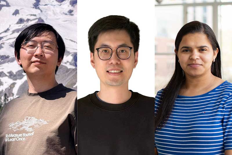
That foundation was the starting point for a recent research project led by Divya Chaudhary, an associate teaching professor at Khoury College in Seattle and director of the Insight X Research Group. Chaudhary, along with computer science master’s students Dong Lu and Yu Zhang, discovered a major problem hiding in the commonly used brain tumor MRI datasets: Many contained duplicated images that could cause AI models to appear far more accurate than they actually were.
Their solution was twofold. First, create a cleaner, more reliable benchmark dataset for brain tumor classification. Second, develop a new AI architecture to improve how modules analyze complex medical images.
The result was “MTA-Swin: A Multi-Token Attention Swin Transformer for Brain Tumor Classification with Leakage-Free MRI Benchmarking,” a paper that introduces both a new data-cleaning pipeline and a new deep learning model that achieved 98.5% accuracy while outperforming 13 representative baseline models.
For Chaudhary, the project began with a simple but important question: Could researchers really trust the impressive results being reported by existing AI systems?
“We found that the digital image libraries used worldwide to train brain tumor AI were full of duplicate images,” Chaudhary said. “AI models looked incredibly smart on paper, but they were actually memorizing leaked images.”
The problem, known as data leakage, happens when information from a model’s training data accidentally appears in its testing data. In medical AI, this can create misleading results. A model may seem capable of diagnosing tumors with near-perfect accuracy, but its performance may not hold up when presented with new patients in a real clinical setting.
Chaudhary compared it to a student taking an exam after accidentally receiving half of the test questions in advance.
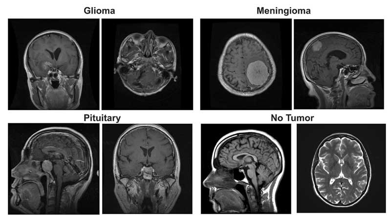
To address this issue, graduate students Lu and Zhang built an automated data-cleaning pipeline that identified and removed duplicate and near-duplicate MRI scans. Using techniques including file fingerprinting and image similarity analysis, the team created a leakage-free benchmark dataset containing 3,522 unique MRI images. The discovery was especially significant because medical imaging datasets are difficult to obtain.
“Because of privacy issues, there are only a few public brain tumor MRI datasets available,” Lu said. “That may be one reason why people overlooked this issue for so long.”
After establishing a cleaner dataset, the researchers turned their attention to improving the AI used to analyze it. Their new model, called MTA-Swin, builds on the existing Swin Transformer, a type of vision transformer commonly used for image analysis. The team modified the architecture by adding a multi-token attention mechanism that allows different parts of the model to exchange information more effectively.
In traditional image-based AI systems, images are often divided into smaller sections for analysis. While this approach can be powerful, it can also cause models to miss subtle details, such as the precise boundaries of a tumor.
MTA-Swin was designed to preserve more of those details.
“Think of it like an expert radiologist,” Chaudhary said. “The model has multiple digital eyes communicating with each other, making sure fine-grained details are not missed.”
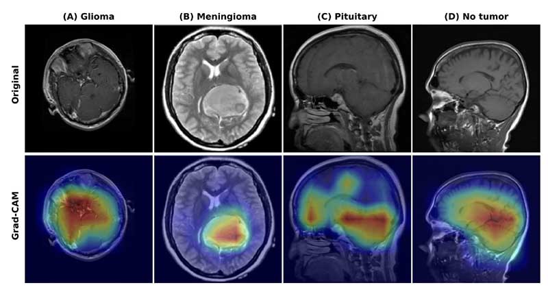
The researchers also focused on making the model more understandable for medical professionals. Using a visualization technique called Grad-CAM, the system can highlight the regions of an MRI scan that influenced its decision, allowing doctors to see whether the AI was focusing on the actual tumor or unrelated image features.
That explainability is critical for health care adoption, Chaudhary said. “Doctors will never trust an AI if they don’t know how it is thinking.”
The model demonstrated several promising capabilities, perfectly identifying healthy brain scans in testing while also showing high precision in detecting aggressive tumors. The system was also designed to operate without requiring expensive GPU infrastructure, potentially making it more accessible for smaller clinics or other resource-limited providers.
Still, the researchers emphasize that AI is not meant to replace physicians.
“AI should not and could not replace doctors,” Zhang said. “It gives doctors a second option. It can help them quickly review MRI images and provide additional information.”
For Zhang and Lu, the project also represented a major milestone as their first published research paper. Both said the experience taught them how to conduct research from the ground up, from reading scientific literature to designing experiments and responding to reviewer feedback.
After developing the initial version of the project in about three months, the team spent several additional months revising experiments and strengthening the paper before it was accepted.
“The research process itself was a very meaningful journey,” Lu said.
Chaudhary credits her students for leading much of the technical work and sees the project as an example of how careful engineering and responsible AI development can improve health care technology.
“The goal is a more reliable, safer clinical diagnosis system,” Chaudhary said. “Not replacing doctors, but giving them better tools to make decisions.”
The Khoury Network: Be in the know
Subscribe now to our monthly newsletter for the latest stories and achievements of our students and faculty